Neuroimage. 2015 May 1;111:350-9. doi: 10.1016/j.neuroimage.2015.02.046. Epub 2015 Feb 28.
Khundrakpam BS1, Tohka J2, Evans AC3; Brain Development Cooperative Group.
CollaboratorsBall WS, Byars AW, Schapiro M, Bommer W, Carr A, German A, Dunn S, Rivkin MJ, Waber D, Mulkern R, Vajapeyam S, Chiverton A, Davis P, Koo J, Marmor J, Mrakotsky C, Robertson R, McAnulty G, Brandt ME, Fletcher JM, Kramer LA, Yang G, McCormack C, Hebert KM, Volero H, Botteron K, McKinstry RC, Warren W, Nishino T, Almli C, Todd R, Constantino J, McCracken JT, Levitt J, Alger J, O’Neil J, Toga A, Asarnow R, Fadale D, Heinichen L, Ireland C, Wang DJ, Moss E, Zimmerman RA, Bintliff B, Bradford R, Newman J, Evans AC, Arnaoutelis R, Pike G, Collins DL, Leonard G, Paus T, Zijdenbos A, Das S, Fonov V, Fu L, Harlap J, Leppert I, Milovan D, Vins D, Zeffiro T, Van Meter J, Lange N, Froimowitz MP, Botteron K, Almli C, Rainey C, Henderson S, Nishino T, Warren W, Edwards JL, Dubois D, Smith K, Singer T, Wilber AA, Pierpaoli C, Basser PJ, Chang LC, Koay CG, Walker L, Freund L, Rumsey J, Baskir L, Stanford L, Sirocco K, Gwinn-Hardy K, Spinella G, McCracken JT, Alger JR,
Author information
- Montreal Neurological Institute, McGill University, Montreal, Canada.
- Electronic address: budha@bic.mni.mcgill.ca.2Department of Bioengineering and Aerospace Engineering, Universidad Carlos III de Madrid, Spain.
- Montreal Neurological Institute, McGill University, Montreal, Canada.
Abstract

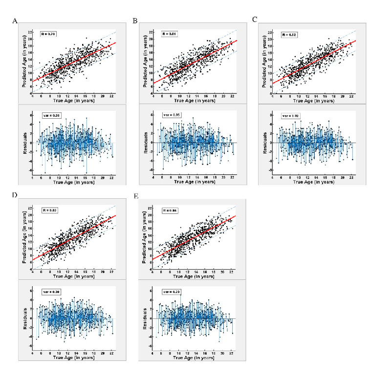
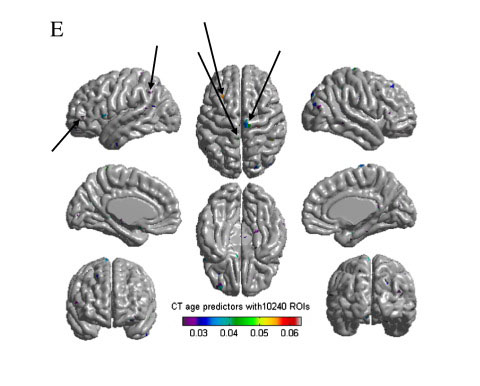
Several studies using magnetic resonance imaging (MRI) scans have shown developmental trajectories of cortical thickness. Cognitive milestones happen concurrently with these structural changes, and a delay in such changes has been implicated in developmental disorders such as attention-deficit/hyperactivity disorder (ADHD). Accurate estimation of individuals’ brain maturity, therefore, is critical in establishing a baseline for normal brain development against which neurodevelopmental disorders can be assessed. In this study, cortical thickness derived from structural magnetic resonance imaging (MRI) scans of a large longitudinal dataset of normally growing children and adolescents (n=308), were used to build a highly accurate predictive model for estimating chronological age (cross-validated correlation up to R=0.84). Unlike previous studies which used kernelized approach in building prediction models, we used an elastic net penalized linear regression model capable of producing a spatially sparse, yet accurate predictive model of chronological age. Upon investigating different scales of cortical parcellation from 78 to 10,240 brain parcels, we observed that the accuracy in estimated age improved with increased spatial scale of brain parcellation, with the best estimations obtained for spatial resolutions consisting of 2560 and 10,240 brain parcels. The top predictors of brain maturity were found in highly localized sensorimotor and association areas. The results of our study demonstrate that cortical thickness can be used to estimate individuals’ brain maturity with high accuracy, and the estimated ages relate to functional and behavioural measures, underscoring the relevance and scope of the study in the understanding of biological maturity.
Copyright © 2015 Elsevier Inc. All rights reserved.


