The Brain Canada Prisma Protocol consists of a resting state fMRI scan, a pair of T1-weighted MPRAGE scans, a T2-weighted scan, a set of diffusion scans, MT-sat scans, and a B1-map. The description of each follows, with notes for the MR technicians.
rs-fMRI
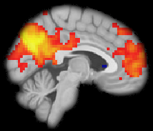
The rs-fMRI scan uses multi-band acceleration to achieve both high temporal and spatial resolution. With an eye to future data sharing, we adopted the ABCD rs-fMRI protocol, with minor adjustments. A slice acceleration factor of 6 allows a TR of 800 ms with 2.4mm iso voxels. A pair of spin-echo scans with opposite phase-encode directions provide for distortion correction. These scans are collected AC-PC -30 (adjusted as need be to fit the brain); the rotation is restricted to the x-axis, i.e. the rotation angles for the y and z axes should be set to 0. The positional parameters from the rs-fMRI should be copied to the distortion correction maps ensuring that the phase encoding directions remain correct. These three sequences should be run back-to-back with no break between scans. The acquisition time for this trio of scans is 7 minutes.
In part, this time is in order to match the play time of the Inscapes movie played during the aquisition. We use a version of the movie that has been scaled down and padded, to reduce the possiblity of the movie inducing saccades. We project video at 1024×768. The screen is placed about 135cm away from the eyes at isocenter and the projected image is about 56cm in width.
Once the three scans for the fMRI data are complete, the Inscapes movie is switched for a real movie (e.g. Finding Nemo), which is shown throughout collection of the structural scans.
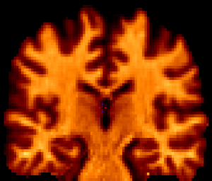
T1
The T1 acquisition comprises a pair of 0.9 mm iso MPRAGE scans. They are accelerated by a factor of 4 as a way to deal with motion. A T1 with no acceleration is about 11 minutes long. The chance of a subject being still for 11 minutes is near zero. The PAT4 scan acquisition time is 3:26; the chance of that being without motion is decent. And, if there is motion, a 3.5 minute scan can be repeated; an 11 minute scan cannot. The acceleration, though, does impact the SNR. SNR drops by roughly the square root of the acceleration factor. That is why there are two T1s. Averaging the two PAT4 T1s returns you to roughly the SNR of a single PAT2 T1, and also reduces the impact of minor motion in either T1. Both T1s are required to be relatively free of motion artifacts. Repeat as necessary.
The T1s are collected with no angles. The position parameters from the first T1 are copied to the second.
T2
The T2 scan is a 0.9 mm iso 3D T2-space. It is, like the T1, accelerated by a factor of 4, but in this case the scanner allowed 2×2 acceleration, which is both faster and yields better SNR. The scan time for the T2 is 3:10, and the quality is sufficient that a single copy is acquired.
The T2 is collected with no angles. The position parameters from the T1s should be copied for the T2.
DWI
The diffusion data are also accelerated, allowing 125 volumes at 1.7 mm iso to be acquired in just over 10 minutes. The diffusion sequences form a 60 direction scan with 4 shells: b=300, b=650, b=1000, and b=2000. The first 7 directions of a standard 60-direction scan are collected with all four b-values. The following 8 directions are collected with b=650, b=1000, and b=2000. The following 15 are collected with b=1000 and b=2000. And the last 30 are collected with only b=2000. So, in total there are 7 directions with b=300, 15 directions with b=650, 30 directions with b=1000, and 60 directions with b=2000.
The acquisition is divided into the 4 sequences described above, thus all b-values in the same direction are acquired back-to-back so that they are likely to in fact be in the same direction, and that moments of motion are more likely to impact a single direction than multiple directions. This also allows an individual sequence to be repeated if motion during acquisition of that sequence is excessive. The acquisition time for each of the sequences is less than 3 minutes.
Thirteen b=0 volumes are distributed across these sequences. An additional sequence collects 7 b=0s in the opposite phase encoding direction to allow distortion correction.
The diffusion scans are collected AC-PC; the rotation is restricted to the x-axis, i.e. the rotation angles for the y and z axes should be set to 0. The positional parameters can be copied from the first sequence to all others, ensuring that the phase encoding directions remain correct. The data should be looked at after each sequence is collected, and if there is too much motion, it should be repeated.
MT-sat
The MT-sat sequences are acquired at the same resolution, i.e. 1.7 mm iso, and in the same space as the diffusion data. This is to facilitate the two modalities being combined to create a map of the myelin g-ratio (the ratio between the inner and the outer diameter of the myelin sheath).
The MT-sat consists of an MToff sequence, an MTon sequence, and a T1-weighted sequence. The acquisition time for the first two sequences is 4 minutes each; the acquisition time for the T1-weighted sequence is 1:50.
The positional parameters from the diffusion sequences should be copied for the MT-sat sequences, ensuring that the phase encoding directions remain correct; but, due to the odd versus even number of slices, the frame must be shifted up 0.85 mm (half of slice thickness).
B1-map
The B1-map scans are a pair of spin echo segmented EPI sequences, one with a flip angle of 60, the other 120. Each has an acquisition time of 1:08.
These are of general use, but are primarily intended to be used in conjunction with the NODDI and MT-sat to derive estimates of the myelin g-ratio.
The orientation from the MT-sat sequences should be copied to the B1-map sequences, ensuring that the phase encoding directions remain correct.