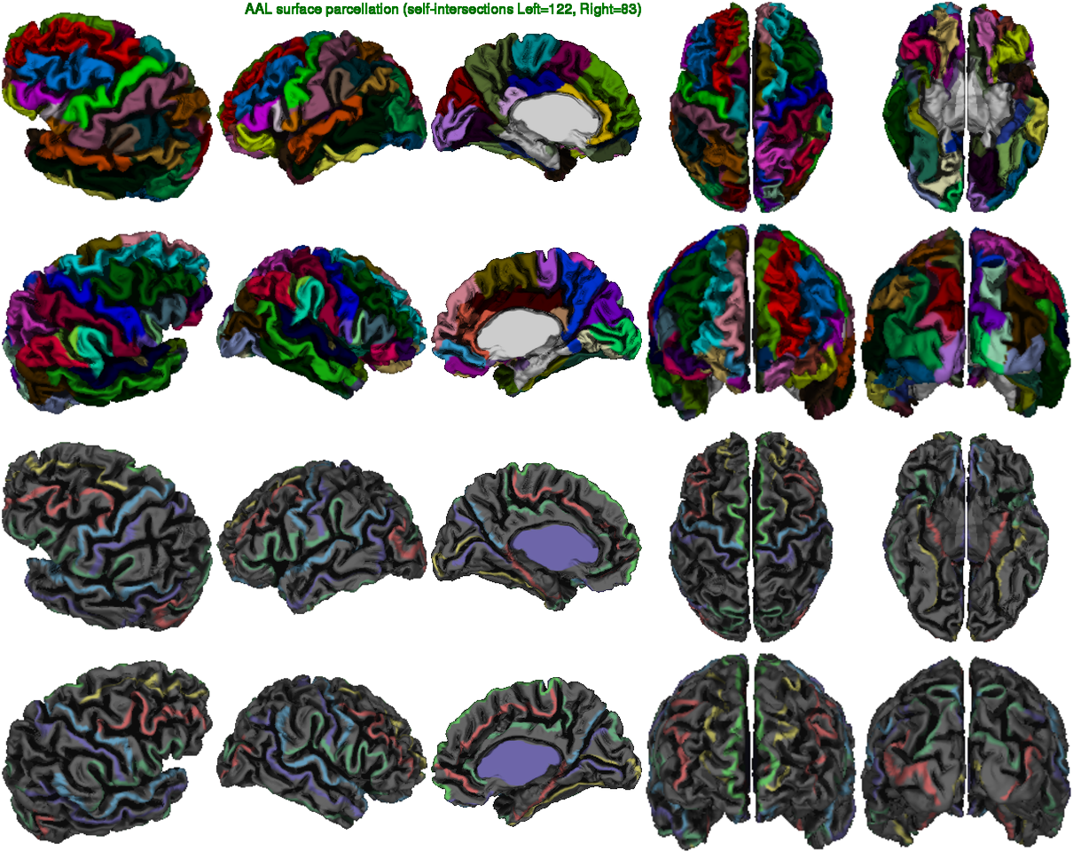
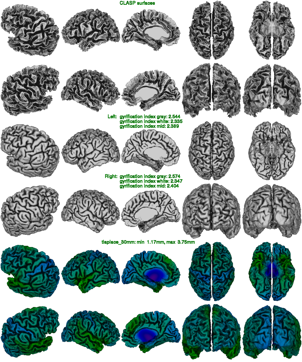
CIVET is an image processing pipeline for fully automated volumetric, corticometric, and morphometric analysis of human brain imaging data (MRI). CIVET performs transformation into stereotaxic space, grey and white matter tissue classification, reconstruction of left and right hemisphere cortical surfaces, surface registration in order to perform group comparisons and cortical thickness analysis. Regional maps are produced based on the lobar parcellation of surfaces. A number of other measurements are also performed, such as mean curvature, gyrification index and total cortical area.
Conversion tools to make comparison between CIVET and other image processing tools are available on demand.
Large-Scale data analysis
The CIVET pipeline can take advantage of distributed compute clusters to expedite preprocessing of large-scale anatomical MRI data through parallel-processing. Features such as automatically-generates quality-control spreadsheets, preprocessing logs, and composite images to provide a visual summary of the pipeline output, facilitate detection of errors and outlying data.
Currently, different versions of CIVET are installed on CBRAIN.
AAL Surface Paracellation
Technical Features of CIVET
CIVET is implemented with high-level scripts, primarily using the common scripting language Perl, that runs computationally efficient image processing tools mainly implemented in C/C++. CIVET extends the previous in-house pipelines by the addition of corticometry analysis tools.
CIVET analysis includes our in-house morphometric analysis tools for:
Boucher M et al (2009) Depth potential function for folding pattern representation, registration and analysis. Med Image Anal [full ref]
Chung MK et al (2005) Unified statistical approach to cortical thickness analysis. Inf Process Med Imaging [full ref]
Chung MK et al (2005) Cortical thickness analysis in autism with heat kernel smoothing. Neuroimage [full ref]
Im K et al (2008) Brain size and cortical structure in the adult human brain. Cereb Cortex [ful ref]
Kabani N et al (2001) Measurement of cortical thickness using an automated 3-D algorithm: a validation study. Neuroimage [full ref]
Kim JS et al (2005) Automated 3-D extraction and evaluation of the inner and outer cortical surfaces using a Laplacian map and partial volume effect classification. Neuroimage [full ref]
Lee JK et al (2006) A novel quantitative cross-validation of different cortical surface reconstruction algorithms using MRI phantom. Neuroimage [full ref]
Lerch JP, Evans AC (2005) Cortical thickness analysis examined through power analysis and a population simulation. Neuroimage [full ref]
Lerch JP et al (2005) Focal decline of cortical thickness in Alzheimer’s disease identified by computational neuroanatomy. Cereb Cortex [full ref]
Lerch JP et al (2006) Mapping anatomical correlations across cerebral cortex (MACACC) using cortical thickness from MRI. Neuroimage [full ref]
Lyttelton O et al (2007) An unbiased iterative group registration template for cortical surface analysis. Neuroimage [full ref]
MacDonald D et al (2000) Automated 3-D extraction of inner and outer surfaces of cerebral cortex from MRI. Neuroimage [full ref]
Robbins S et al (2004) Tuning and comparing spatial normalization methods. Med Image Anal [full ref]