Cereb Cortex. 2017 Feb 18:1-11. doi: 10.1093/cercor/bhx038. [Epub ahead of print]
Khundrakpam BS1, Lewis JD1, Kostopoulos P1, Carbonell F1, Evans AC1.
Author information
- Montreal Neurological Institute, McGill University, Montreal, QC, Canada H3H2P1.
Abstract

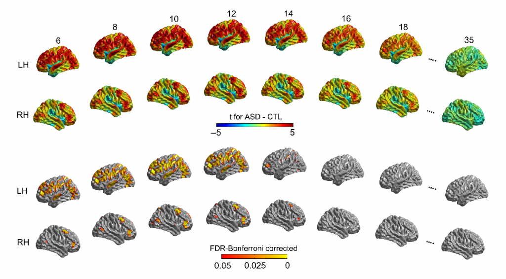
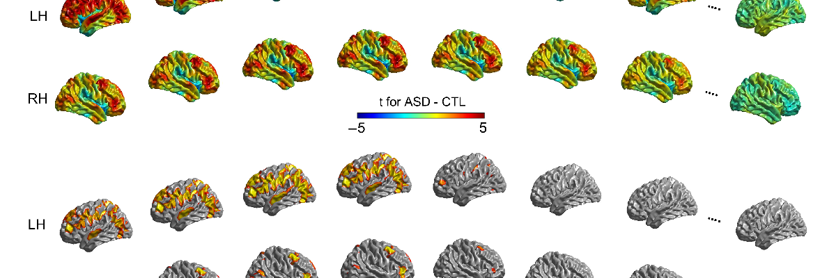
Neuroimaging studies in autism spectrum disorders (ASDs) have provided inconsistent evidence of cortical abnormality. This is probably due to the small sample sizes used in most studies, and important differences in sample characteristics, particularly age, as well as to the heterogeneity of the disorder. To address these issues, we assessed abnormalities in ASD within the Autism Brain Imaging Data Exchange data set, which comprises data from approximately 1100 individuals (~6-55 years). A subset of these data that met stringent quality control and inclusion criteria (560 male subjects; 266 ASD; age = 6-35 years) were used to compute age-specific differences in cortical thickness in ASD and the relationship of any such differences to symptom severity of ASD. Our results show widespread increased cortical thickness in ASD, primarily left lateralized, from 6 years onwards, with differences diminishing during adulthood. The severity of symptoms related to social affect and communication correlated with these cortical abnormalities. These results are consistent with the conjecture that developmental patterns of cortical thickness abnormalities reflect delayed cortical maturation and highlight the dynamic nature of morphological abnormalities in ASD.
© The Author 2017. Published by Oxford University Press.For Permissions, please e-mail: journals.permissions@oup.com.
PMID:28334080 | DOI:10.1093/cercor/bhx038


